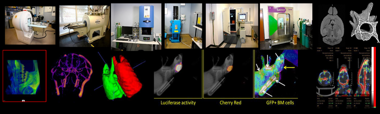
Small Animal Imaging
The Small Animal Imaging Resource provides a wide range of imaging and radiation treatment resources for animal research. The preclinical services are translational in operation while remaining cutting edge in the advancement of basic science research. Services are available to the Augusta University research enterprise and other outside scientific investigators.
Image Quality Assurance & Project Management
The Small Animal Imaging shared resource takes an orderly approach to managing your imaging project and employs a regimented method for providing Quality Assurance and Control for all image acquisition and image analysis. Small Animal Imaging shared resource incorporates years of experience in research project management and preclinical level image analysis in every imaging project that it is involved for translational studies.
The Small Animal Imaging shared resource provides low-cost imaging services to the Augusta University research community. Prices for imaging, data analysis, and consultation are intended to be competitive with the charges for similar services at other academic institutions.
Contact Us
Small Animal Imaging
Health Sciences Campus
GCC - M. Bert Storey Research Building
CN 1153, 1188, 2144, 5162
Asamoah Bosomtwi, PhD
Director
CN-3155
(706) 721-1427
Images
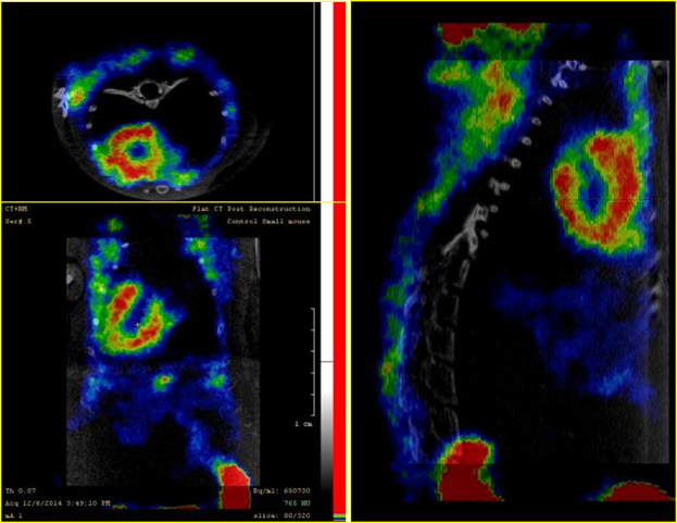
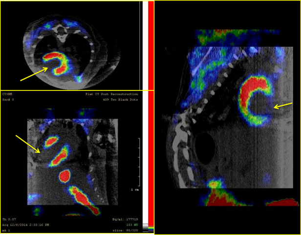
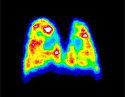
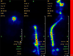
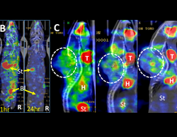
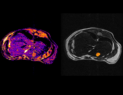
Single photon emission computed tomography (SPECT)
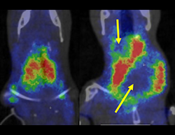
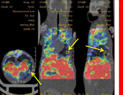
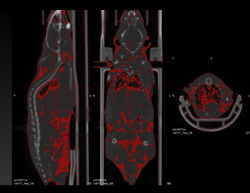
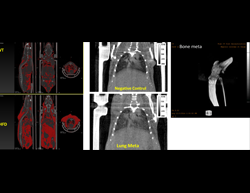
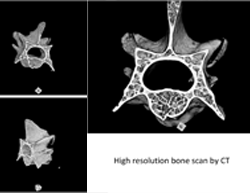
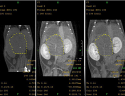
Computed topography
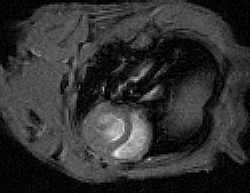
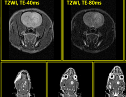
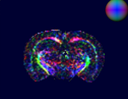
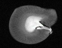
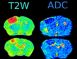
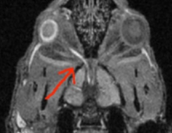
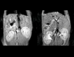
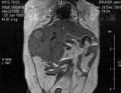
MRI Images
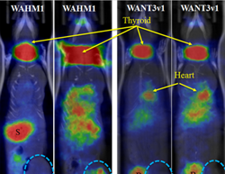
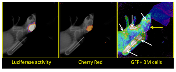
Bioluminescence Imaging/Fluorescence Imaging
Fee & Payment
Small Animal Imaging provides low-cost imaging services to the Augusta University research community.
Prices for imaging, data analysis, and consultation are intended to be competitive with the charges for similar services at other academic institutions. All charged usage will be pro-rated in ½-hour blocks, in accordance to time used. All scheduling and billing are through iLab.
Equipment
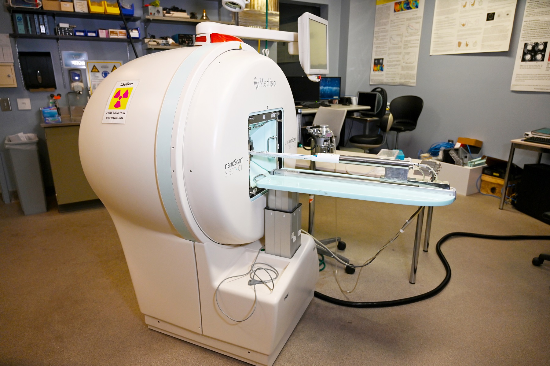
Mediso SPECT/CT System
Single-photon emission computed tomography (SPECT)is a nuclear medicine tomographic imaging technique using gamma rays and provides 3D images of radiotracer distribution. It is very similar to converting nuclear medicine planar imaging using averaging camera. Combine with computed tomography (CT), anatomic localization can be identified. Traditional radiotracers or novel proteins and antibodies of interest that can be radiolabeled with gamma emitting isotopes, injected into subjects, and imaged to determine organ and/or tumor specificity.
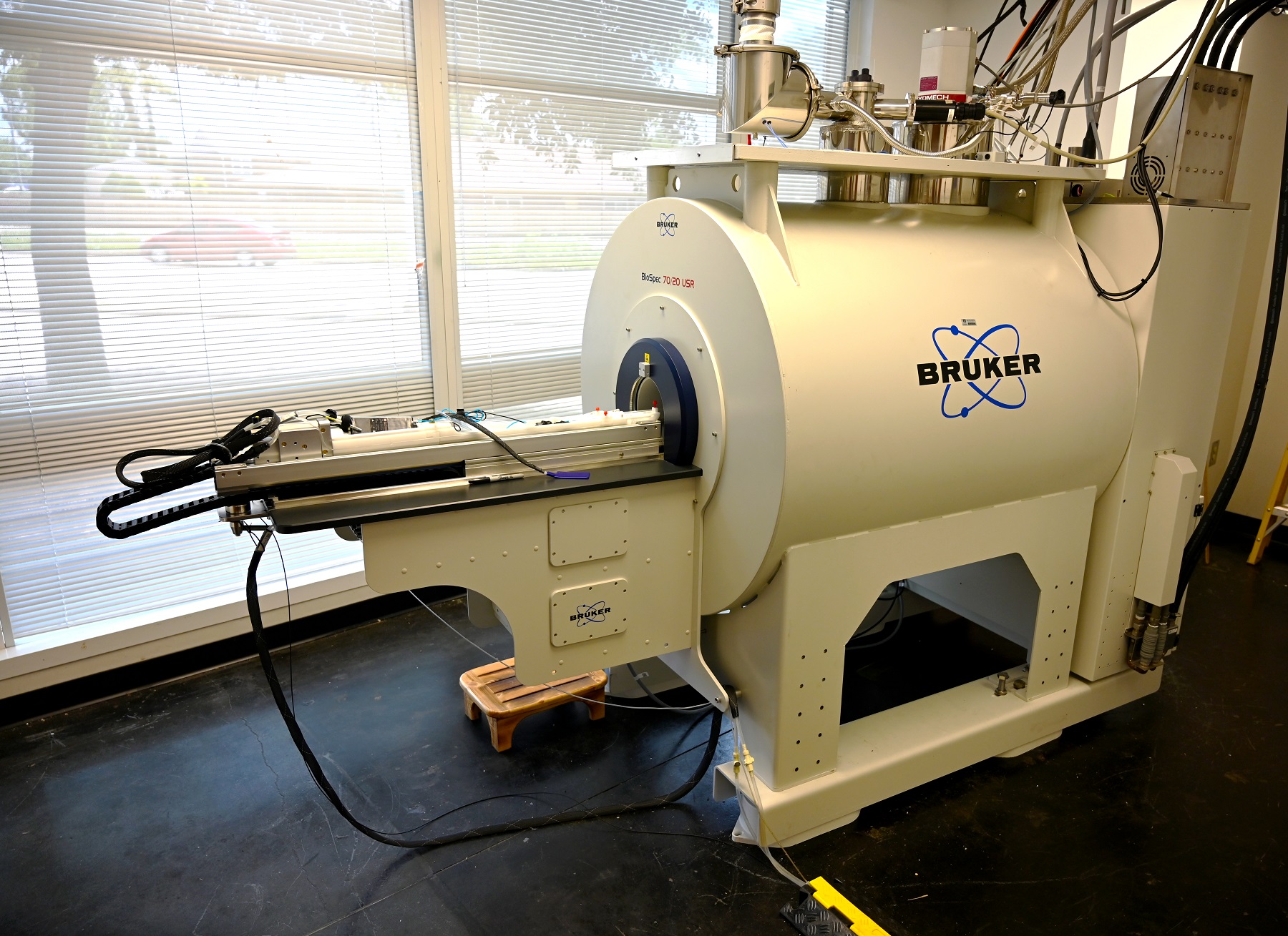
7T Bruker Biospin Magnetic resonance Imaging System
MR imaging is a method for non-invasive imaging that can provide information related to regions of interest and their surrounding environment. MRI can be used to find problems such as tumors, bleeding, injury, blood vessel diseases, or infection. Basic and specialized scans can be performed on the 7T MRI located in CN-1153
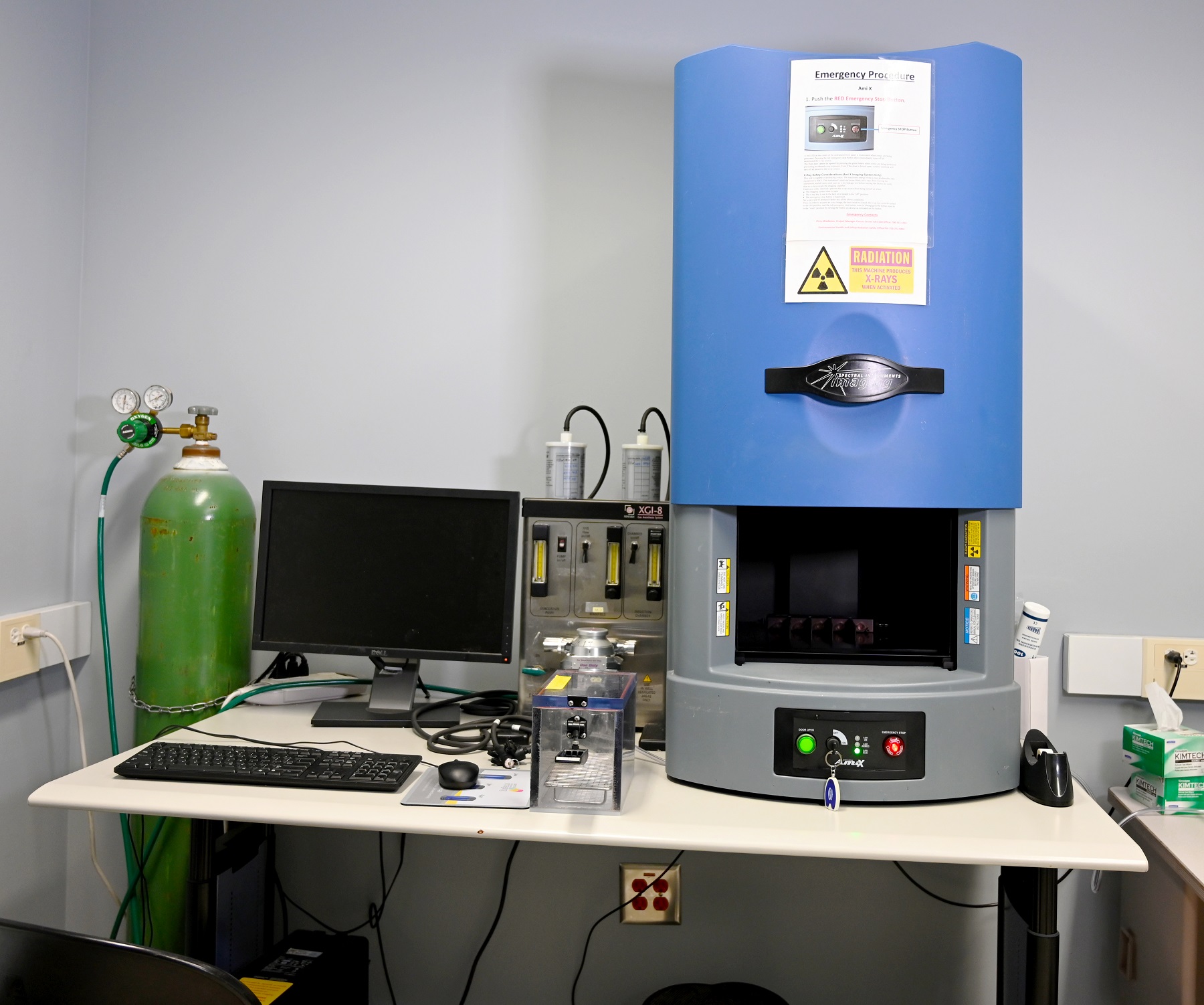
Spectral AMI X Bioluminescent/Fluorescent Imaging System
Bioluminescent and fluorescent optical images are created non-invasively by detecting the photons from tracers that safely emit from areas of interest in the animal or cell lines. The sensitivity of the cameras is such that the optical imager can detect wavelengths from 400 to 800 nm and from source sizes as small as four to five cells.
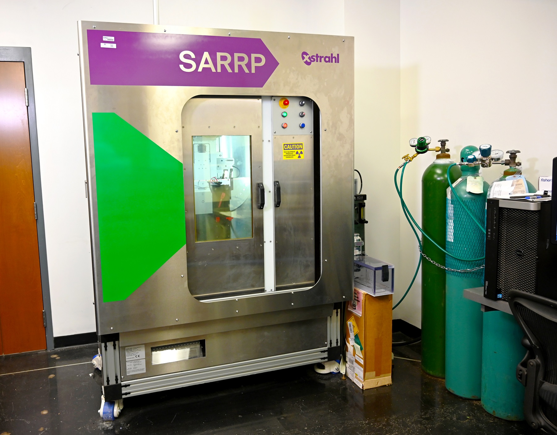
Small Animal Radiation Research Platform
The Small Animal Radiation Research Platform (SARRP) is to make the animal models available for the development and evaluation of novel radiation therapies. In combination with advanced imaging methods, small animal research allows detailed study of biological processes, disease progression, and response to therapy, with the potential to provide a natural bridge to the clinical environment.

Precision X-RAD 320
The Precision X-Rad 320 offers a large chamber within a compact cabinet footprint. It is a self-contained x-ray system designed to deliver a precise radiation dosage to specimens from cells to small animals such as rabbits. The x-ray tube is used specifically for radiation therapy, having a highly homogenous beam.Featuring our TouchRad control panel - a multi-user, password protected touchscreen interface - the X-Rad320 includes a transportable database that can track individual system usage.
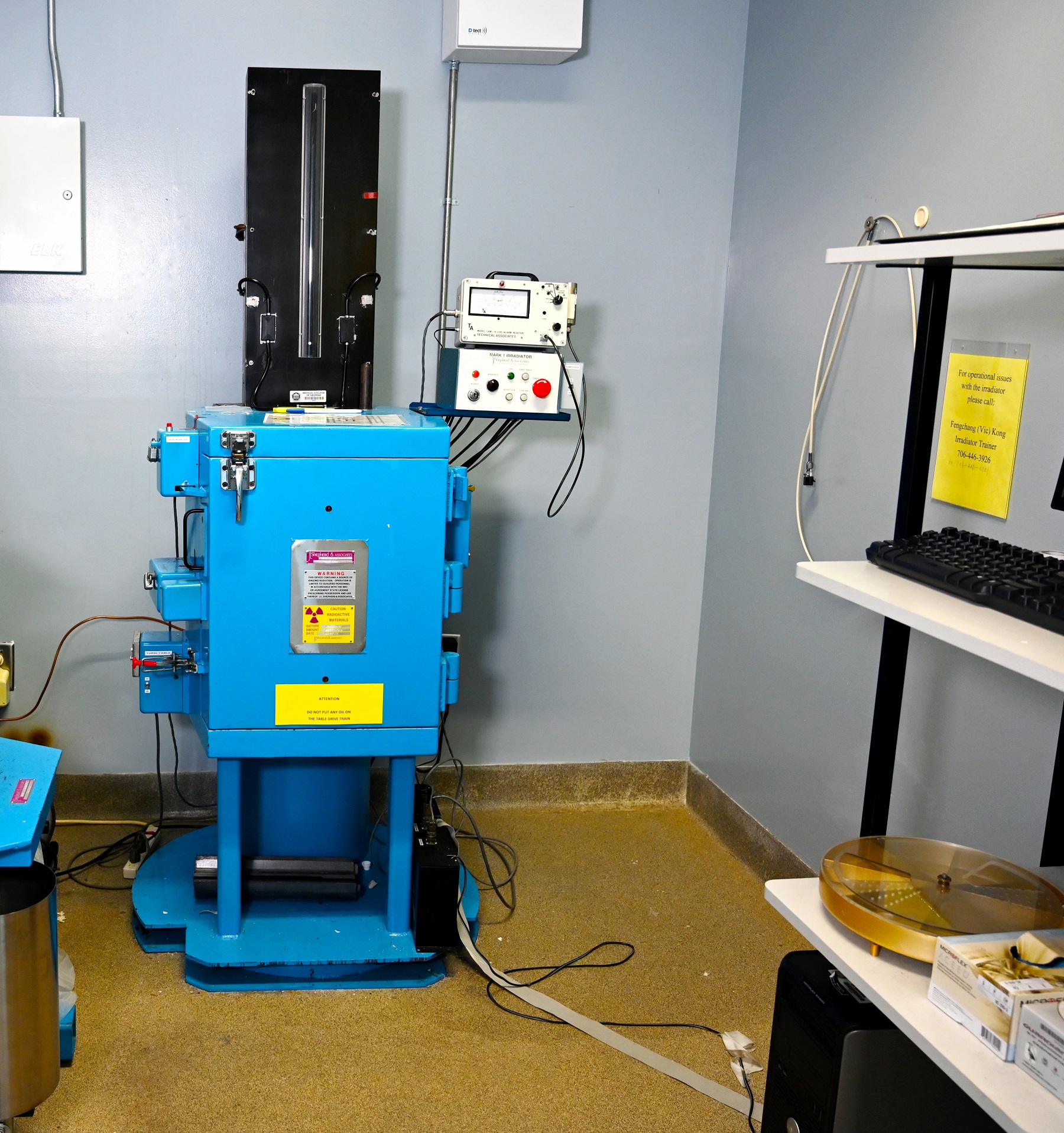
JLS Gamma Cell Irradiator
JLS Mark I Irradiators irradiate samples on turntables adjacent to a line source of Cs137. This provides high radiation levels for irradiating cells/tissue cultures in test tubes/culture vessels ≤ 100 mm diameter and lower dose rates to large numbers of mice/rats in animal chambers on large turntables at a greater distance from the Cs137 source in the same irradiator.
Meet Our Team


Elizabeth Rodgers