- Augusta University
- Colleges & Schools
- Medical College of Georgia
- Dermatology
- Mohs Micrographic Surgery
Mohs Micrographic Surgery
Mohs micrographic surgery is a procedure performed at Augusta University Medical Center by Dr. Matthew Belcher, a fellowship trained Mohs micrographic surgeon and board certified dermatologist.
Mohs micrographic surgery is the most effective way of removing certain skin cancers, particularly those that spread deep and wide with little finger-like or root-like projections. This kind of tumor spread is called contiguous or direct spread and differs from metastatic spread by invading only local tissues and not distant sites such as lungs and liver. Contiguous spread pushes normal tissue out of the way or simply destroys it. Tumors that spread this way include basal cell and squamous cell carcinoma. There are also a large variety of related skin tumors called appendageal tumors which can spread contiguously. These types of tumors are all capable of being treated by Mohs micrographic surgery (or simply Mohs surgery, or just "Mohs").
Mohs surgery is named for Frederick Mohs, the physician who developed the technique. At first, the surgery required putting several chemicals on the skin to "fix" or mummify the area involved with tumor prior to the surgery. These chemicals were painful and needed to be affixed for about half a day. The advantage was that the surgery could take place without anesthesia and with no bleeding. When the technique was first introduced it was called chemosurgery because of the chemicals used. But, the technique has been modified significantly since Dr. Mohs first described it, and the chemicals are virtually never or extremely rarely ever used anymore.
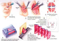
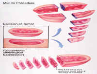
Mohs surgery is different from conventional surgery in several very special ways. After debulking, the area involved with visible tumor is removed surgically in a thin beveled disc, very much like a coin. This is done under local anesthesia and after a drawing of the involved area has been made. The skin is scored to help maintain proper orientation and positioning.
 Once removed, the disc-shaped piece of tissue is cut into smaller pie-shaped pieces.
Each piece is numbered and color-coded with several dyes so that the doctor can identify
exactly where each piece came from and how it was oriented on the skin. All of this
information is transferred to the drawing which was made of the area, and the tissue
is taken to the lab for immediate processing. While the tissue is being processed,
the patient's wound is dressed, and the patient can wait comfortably in the surgical
room or in the surgery or main waiting rooms. Processing can take about thirty minutes
for an average amount of tissue but may take longer for larger amounts of tissue.
 Processing is extremely important as it helps to identify any areas were tumor might
have been left behind. During the processing, the tissue is compressed and frozen
rapidly with liquid nitrogen. The tumor is then cut into extremely thin sheets from
the bottom surface up, with a special machine called a cryostat. These thin sheets
of tissue are then put on a microscope slide and stained. Once the staining is done,
the final touches are put on the slide and it's ready for the doctor to look at under
the microscope.
What the doctor will look at under the microscope represents all of the bottom and all of the edges of the original disc of tissue. since the tumors being dealt with spread deep and wide, the doctor is looking at all possible areas where tumor could still be present. This is much more accurate than conventional methods.
If any tumor is present, the doctor can identify it and its exact location because of the color-coding which was done immediately after the piece was removed surgically. The information is transferred to the drawing which will then serve as a map for the doctor and the rest of the surgical team. In this way, the exact location of the tumor can be identified for the second stage of surgery. Obviously, if all the tumor was removed in the first stage, no additional surgery will be necessary. This is quite often the case.
 If a second stage of Mohs surgery is necessary, only those areas that are positive
for tumor will be removed. This leaves in place as much normal tissue as possible.
All other surgical steps, including color-coding, processing and microscopic evaluation
are the repeated. The entire process is repeated as often as necessary as long as
tumor is still present. Mohs surgery is completed when no more tumor is present in
the tissue examined under the microscope. when Mohs surgery is completed, there are
no more microscopic projections of tumor remaining. Additionally, as much normal and
healthy tissue has been left in place.
Once the Mohs portion of the surgery is done, a defect or hole will be left where the tumor once was. Often times this can be repaired quite simply. Occasionally, a more complicated repair is necessary and this might mean the use of a skin graft or flap. Rarely, when a repair becomes extremely complicated, it may require that the surgery be done in stages or perhaps by other physician specialists. This may mean a short stay in the hospital and surgery under general anesthesia. Fortunately, the great majority of repairs are simple and are outpatient procedures.
Fairly often, rather than a repair, a wound may be left to heal by itself. This is called second intention healing or healing by granulation. This type of healing may be chosen over repair because the cosmetic result is expected to be better or because it will heal into a smaller scar which can be more easily repaired at a later date. When a wound, even a large one is left to heal by second intention, the wound is usually remarkably pain-free! There may be other reasons for selecting second intention healing and these would be discussed by the doctor.
Skin cancer surgery has several objectives or priorities. The first is always to remove all the tumor. Once this has been accomplished, the next priority is cosmetic appearance. The doctor will always discuss the various options and rationale for repair or second intention healing after the tumor has been removed. When repairs are uncomplicated they will usually be done immediately while the area is still numb.
Not all skin cancers require Mohs surgery. The procedure is usually selected for tumors growing in high-risk areas of the face such as the nose, eyes, mouth, ears and surrounding tissue. It is also selected for all recurrent tumors (those reappearing after having been removed), all large tumors and all tumors which look aggressive under the microscope. Your doctor will advise you on the technique that is appropriate for your particular condition.
A limited number of doctors are qualified to perform Mohs surgery. These are Dermatologists who have completed an approved training fellowship in Mohs surgery and who are members of the American College of Mohs Micrographic Surgery and Cutaneous Oncology. Members of this organization are recognized as fully competent to perform and teach this procedure. When properly done, this technique has the highest cure rates possible, often approaching 100%.

Matthew Belcher, MD