Back to Top
Apply Giving myAugusta
Resources for:
Students
Students Faculty & Staff Parents & Family Alumni Community
Augusta University Logo
  • Academics
    Graduates pose together in their regalia.
    Augusta University celebrates fall 2025 graduates »

    Academics Home

    Colleges & Schools

    Programs of Study
    • Degrees & Programs
    • Course Catalog
    • Course Schedule
    • Program Pathways
    • Academic Calendar
    • Online Programs
    • Accelerated Degree Programs
    Resources
    • Academic Success Center
    • Advising
    • Counseling Services
    • Honors Program
    • Libraries
    • Testing & Disability Services
    • Writing Center
    Outside the Classroom
    • Army ROTC
    • Study Abroad
    • Experiential Learning
    • First Year Experience
    • Center for Undergraduate Research
    • Career Services
    • Jags Live Well
  • Admissions
    Augusta University's jaguar mascot, Augustus, and two students hold their hands like a paw
    Augusta University awarded $1.3 million grant to expand student support »

    Admissions Home

    Visit Campus

    Request Information

    Apply to AU
    • First-Year Freshmen
    • Transfer Students
    • Dual Enrollment
    • Graduate Students
    • Medical College of Georgia
    • Dental College of Georgia
    Opportunities
    • Degree & Programs
    • Honors Program
    • Program Pathways
    • Military-Affiliated Students
    • New Student & Family Transistions
    • On-Campus Housing
    Financial Aid
    • Student Financial Aid
    • Net Price Calculator
    • Scholarships
    • Cost of Attendance
    • Apply for Federal Aid
  • Campus Life
    Two students pose with the university mascot, Augustus, showing their Jaguar pride.
    Augusta University awarded $1.3 million grant to expand student support »

    Campus Life Home

    Athletics

    Community
    • Army ROTC
    • Living-Learning Communities
    • Military & Veteran Services
    • Mentorship
    • New Student & Family Transistions
    • Jags 4 Jags Mentoring Program
    Campus Services
    • Dining Services
    • Roarstore
    • Housing
    • Student Health
    • Parking & Transportation
    • Jagcard
    Get Involved
    • Clubs & Organizations
    • Greek Life
    • Campus Recreation
    • Student Government
    • Jaguar Production Crew
    • Intramural Sports
  • Research
    Three men in suits stand in front of a Augusta University Medical College of Georgia backdrop and smile at the camera. The man in the middle is holding a plaque.
    MCG scientists investigate arthritis drug’s impact on Alzheimer’s disease »

    Research Home

    Opportunities
    • Undergraduate Research
    • Graduate & Postdoctoral Research
    • Clinical Trials
    • Core Laboratories
    • Innovation Commercialization
    Initiatives
    • Cancer
    • Cardiovascular
    • Immunology
    • Neuroscience
    • Aging
    Resources
    • Centers & Institutes
    • Ethics & Compliance
    • Institutional Review Board
    • Sponsored Programs
    • Tools for Researchers
  • About AU
    Five women and two men jump and raise their hands in celebration.
    AU contributed over $1.6 billion to Georgia’s economy in FY 2024 »

    About AU

    Jagwire News

    Leadership
    • President
    • Provost
    • Administration
    • Enrollment Student Affairs
    • Faculty Senate
    We are AU!
    • Our Mission
    • Working at AU
    • Traditions
    • History
    • Augusta, GA
    Resources
    • MyAugusta
    • Calendar of Events
    • Brand Guidelines
    • Portals
    • Faculty Directory
Resources For
  • Current Students
  • Faculty & Staff
  • Parents & Family
  • Alumni & Friends
Apply
Giving
MyAugusta
Trending Search Terms
  • D2L LMS
  • Email
  • Pounce
  • Calendar
  • Registrar
  • Housing
  • Academic Calendar
  • Financial Aid
  • Parking
  • Library
  • Human Resources
  • Information Technology
Medical College of GeorgiaRadiology & Imaging
Radiology & Imaging
  • People
    • Welcome from the Chair
    • Our Faculty
    • Our Residents & Fellows
    • Past Graduates
  • Education
    • Diagnostic Radiology Residency Program
    • Integrated Interventional Radiology Residency Program
    • Prospective Resident Application Info
    • Curriculum / Call Schedule
    • Life in Augusta
    • Research
    • Benefits
    • Breast Imaging Fellowship Program
    • Neuroradiology Fellowship Program
    • Diagnostic Medical Physics
    • Resident Resources
  • Patient Information
  • Equipment
  • Augusta University
  • Colleges & Schools
  • Medical College of Georgia
  • Radiology & Imaging
  • AMRI

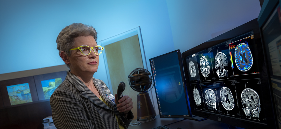
Advanced Clinical Neuro MR Imaging

MCG is the only medical center in the CSRA (Central Savannah River Area) to offer a variety of advanced magnetic resonance imaging.

Advanced MR Imaging differs from typical MRI, which only provides structural information regarding organs such as the brain, spinal cord, and various structures of the head and neck. Physicians specializing in the diagnosis of neurological diseases with high-tech imaging (neuroradiologists) oversee the performance of these special studies which are generally only available at major academic medical centers in the United States such as the Georgia Health Sciences University. Here is a brief description of these techniques and a few examples of clinical applications for each:

FUNCTIONAL MRI-This is a method of evaluating regional changes in blood flow associated with various types of brain activity. Each brain activity is associated with a task that one may perform in everyday life. Such activity may include moving a finger, think of words that rhyme, or remembering a set of numbers. By observing regional changes in blood flow and analyzing data from these images, maps of activation can be generated which display the active areas in the brain responsible for these various important functions (e.g., movement, speech, emotions, and memory). This method has been used by neuroradiologists to aid neurosurgeons in treatment planning prior to surgical removal of brain tumors, vascular malformations, cortical malformations and other intracranial mass lesions in order to avoid surgical removal of vital brain tissue involved in these important functions and thus reduce overall surgical risk.

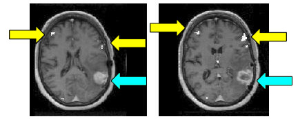
Example of a language task with fMRI showing activation (yellow arrows) far removed from an enhancing tumor (blue arrows):
image 007

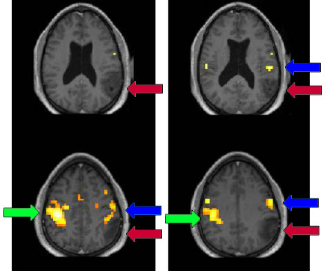
Example of a patient with a left parietal tumor (red arrows) performing a left hand motor task; primary right motor cortical activation (green arrows) is seen, but displacement of left hemispheric activation (blue arrows) is also demonstrated. Some of the left hemispheric activation borders on the margins of the tumor. Knowing this will help a neurosurgeon decide how much of a tumor can be safely removed without causing a postoperative permanent neurological deficit (e.g., paralysis, numbness, or difficulty with movement).

image 019

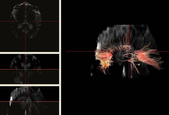
DIFFUSION TENSOR IMAGING -This new method allows neuroradiologists to visualize and characterize the health and direction of white matter tracts in the brain. One can think of these tracts as "wiring" between different parts of the brain that allow us to perform different tasks. If this "wiring" breaks down, as in the case of a stroke or an invasive tumor, brain behavior may be affected. This technique is very useful in diseases such as multiple sclerosis. Variations of this diffusion technique have recently been  commonly used to diagnose acute strokes, but the ability to closely examine white matter in the brain is relatively new.

image 021

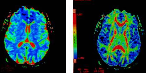
Apparent diffusion coefficient (ADC) map on the left and fractional anisotropy (FA) map on the right of a normal volunteer, demonstrating normal white matter tracts in the brain (on the color scale, red regions are higher in [ADC or FA, respectively] value than blue regions).

In addition, diffusion tensor imaging allows tractography, enables visualization of white matter tractsas they appear in the brain, as demonstrated below (displaying the genu and splenium of the corpus callosum):

image 023

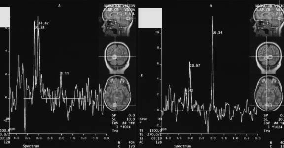
MAGNETIC RESONANCE SPECTROSCOPY ---This is a method to examine the brain metabolites to evaluate the nature of brain mass lesions which may be nonspecific in appearance on conventional structural MRI. This is often used to confirm absence or presence of tumor, although other applications also exist.

The following proton magnetic resonance spectroscopy study demonstrates a right occipital lobe tumor, with associated typical metabolic abnormalities (reduction in NAA and slightly elevated choline peak); the normal left occipital lobe spectrum is on the right, and the abnormal tumor spectrum is on the left side of the image below:

image 025

MCG currently has two 1.5 tesla MRI scanners in addition to a third in an adjacent affiliated VA hospital, and a new state-of-the-art 3.0 T MRI scanner.

We are involved in multiple research projects at MCG involving advanced MR imaging as well, involving multiple sclerosis, brain tumors, and epilepsy.

University Shield

Augusta University

1120 15th Street, Augusta, GA 30912

  •   Campus Maps
  •   Campus Contacts
  • A-Z Directory
  • Degrees & Programs
  • Employment
  • Accessibility
  • Accreditation
  • Campus Safety
  • Compliance Hotline
  • Human Trafficking Notice
  • Privacy Notices
  • Title IX / Sexual Misconduct
Apply Now Give Now

© 2025 Augusta University

Facebook Twitter LinkedIn Youtube Instagram
©