Human MRI Imaging Core
The Human MRI Imaging Core Facility is a core dedicated to providing cutting-edge magnetic resonance imaging services for human participants.
- Current Instruments
- About Us
- Neuroimaging
- Abdominal Imaging
- MSK Imaging
- Vascular Imaging
- Cardiac Imaging
- MR Spectroscopy
- Breast Imaging
- Other Imaging
- Advisory Committee
Policy and price for using 3T Research MRI
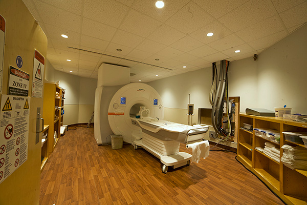
Instruments
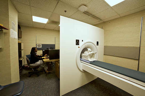
Siemens Vida 3T MRI Scanner
- Technical operation and safe work environment provided by clinically certified MR technologists.
- Equipped with high-performance imaging coils (e.g., 64-channel head coil), for high-resolution and accelerated imaging.
- Paired with functional MRI stimulation hardware and software from Nordic Neuro Lab.
- Capable of performing a wide range of clinical and research techniques, including:
- High-resolution structural imaging (e.g., MSK, Brain)
- Spectroscopy
- Task-based, event-based, and resting-state Functional MRI (fMRI)
- Perfusion
- Anisotropic Diffusion Imaging
- Relaxometric parameter mapping
- MR Elastography
PST Mock MRI Simulator
- Participant acclimation in the MRI environment (e.g., Pediatric studies)
- Reduction of failed scans from participant motion or claustrophobia
- Training of participant for functional imaging tasks
3T Siemens Magnetom Vida with BioMatrix technology and turbo suite excelerate package
- Features:
- Bore size – 70 cm
- Gradient amplitudes: 60mT/m
- Maximum Gradient slew rate: 200 T/m/s
- Maximum number of RF receivers : 128 independent channels
- Minimum FOV 5 mm
- Max FOV 55x55x50 cm
- Coils available:
- 64 channel head
- 20 channel head and neck
- 32 channel spine (posterior)
- 18 channel standard body
- 18 channel large & small flex
Imaging
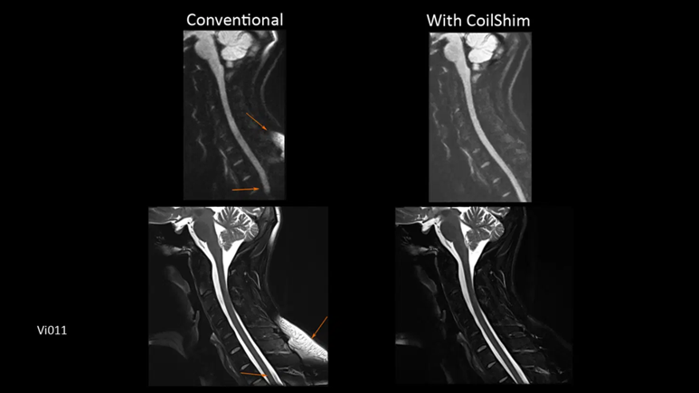
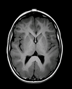
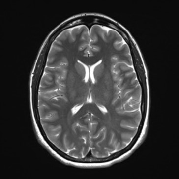
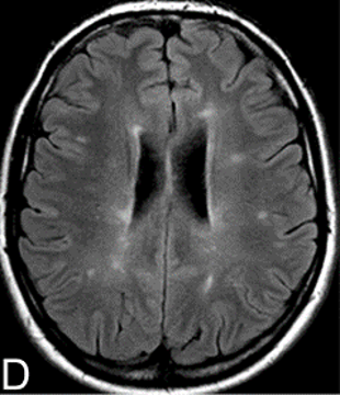
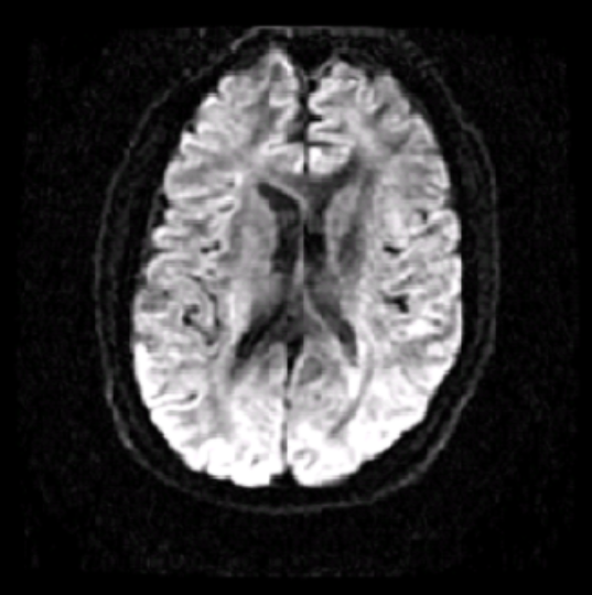
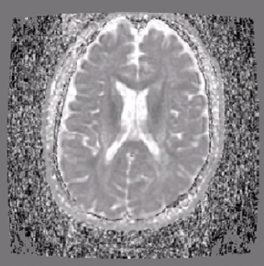
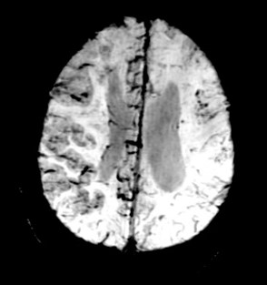
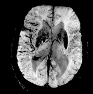
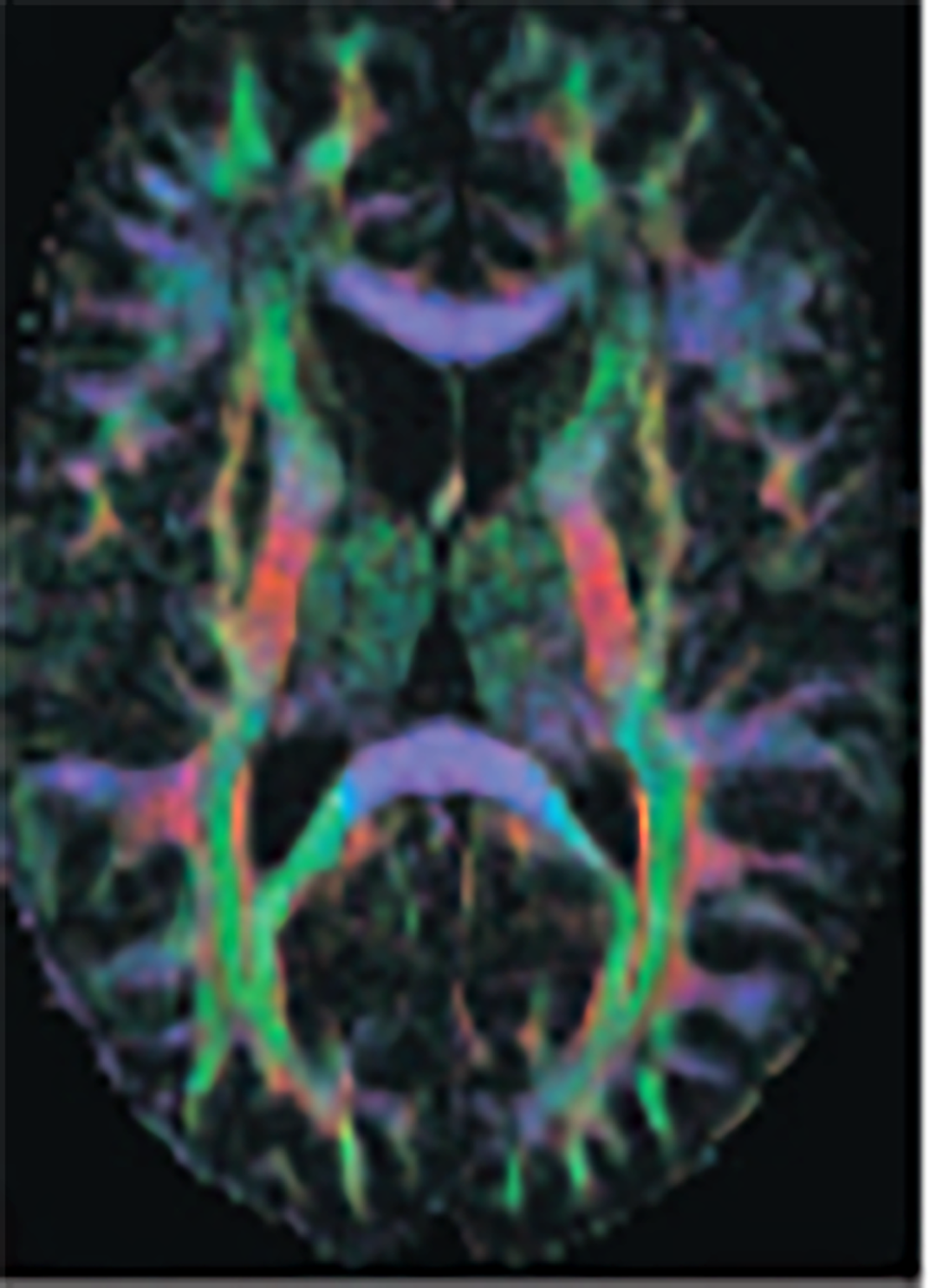
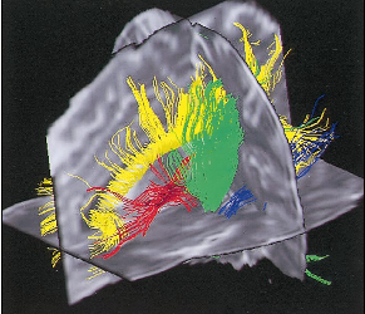
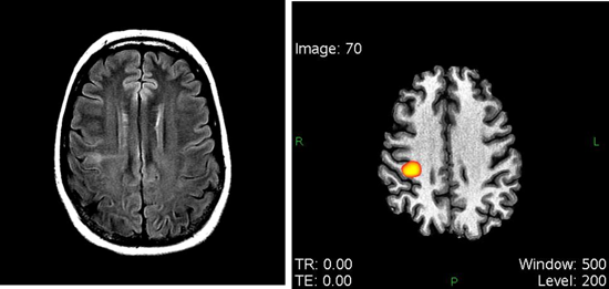
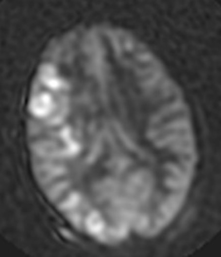
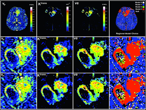
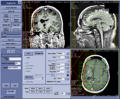
Neuroimaging
All basic MRI for anatomical images (like T2WI, T1WI, GRE, etc), Neuro perfusion, DSC-based perfusion, DCE-based perfusion and permeability, Cerebral blood flow (2D and 3D ASL), Advanced less-distorted DWI, Selective excitement. DTI (B-value ranges from 0 -10000 s/mm2, directions 256) and fMRI (NordicNeuroLab fMRI and Visual system).
Click images for larger view and description

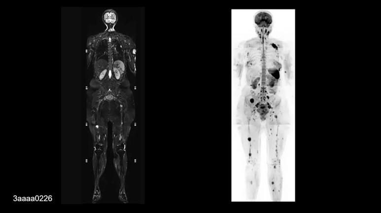
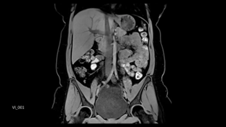
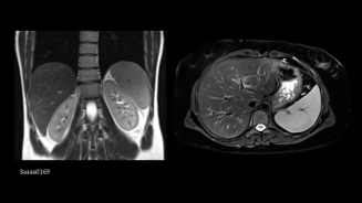
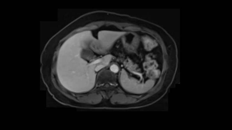
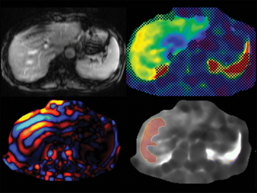
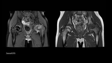
Abdominal Imaging
All standard sequences for anatomical imaging with or without fat suppression. All advanced sequences for liver imaging for determining fat and iron, MR Elastrography to determine liver fibrosis.
Click images for larger view and description
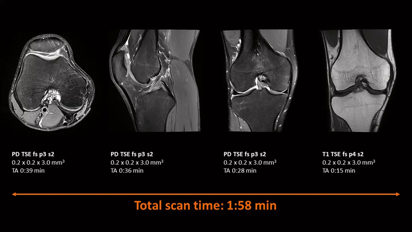
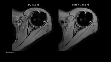
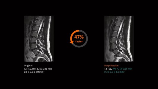
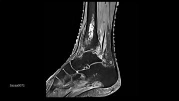
MSK
Basic sequences, T1, T2, T2* mapping. WARP and Advanced WARP (metal implant).
Click images for larger view and description
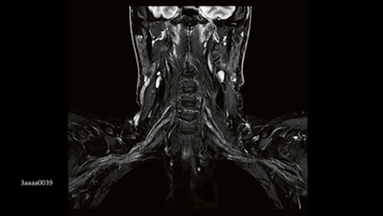
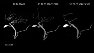
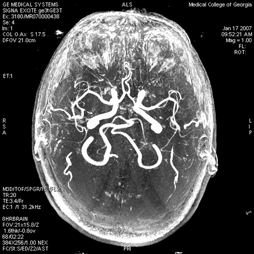
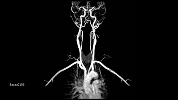
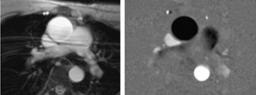
Vascular Imaging
Pre and post-contrast MRA. Post-contrast multiphasic MRA. MR-DSA. Arterial wall thickness and atherosclerotic plaques can be determined.
Click images for larger view and description
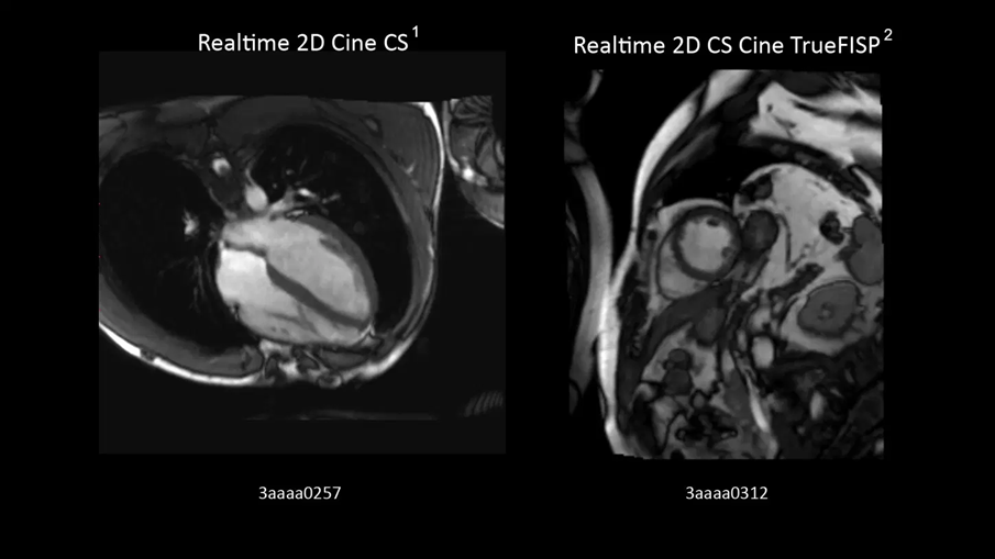
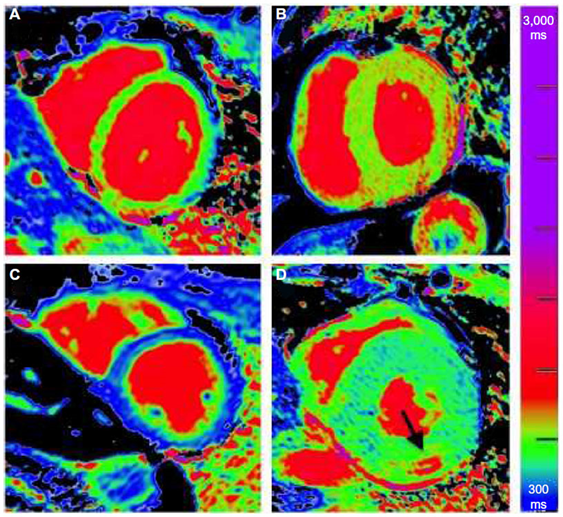
Cardiac Imaging
MRI is considered gold standard for myocardial thickness and changes in the relaxivity due to chemotherapy and other injury. T1- T2-, T2* maps can be created.
Click images for larger view and description
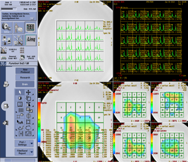
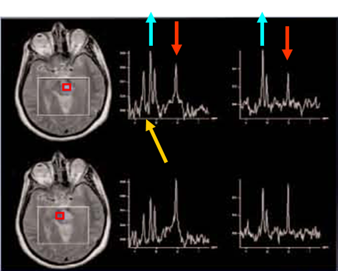
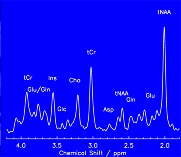
Magnetic Resonance (MR) Spectroscopy
Magnetic resonance (MR) spectroscopy is a noninvasive diagnostic test for measuring biochemical changes in the brain, especially the presence of tumors. MR spectroscopy compares the chemical composition of normal brain tissue with abnormal tumor tissue.
Click images for larger view and description
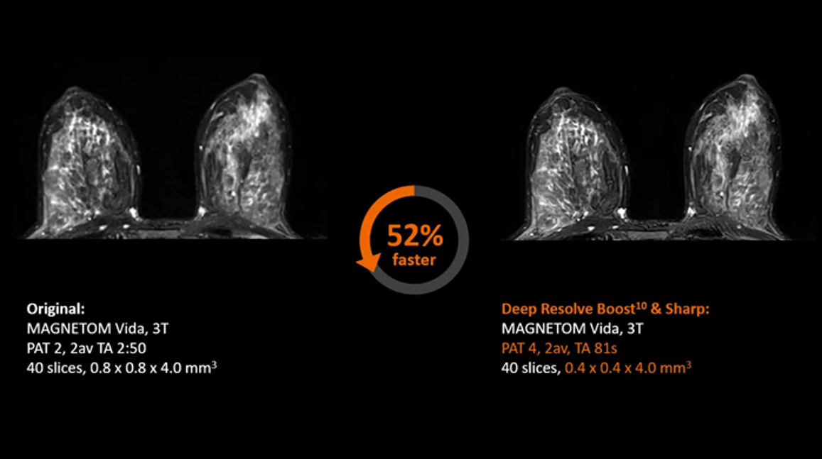
Breast Imaging
Other Imaging
Click images for larger view and description